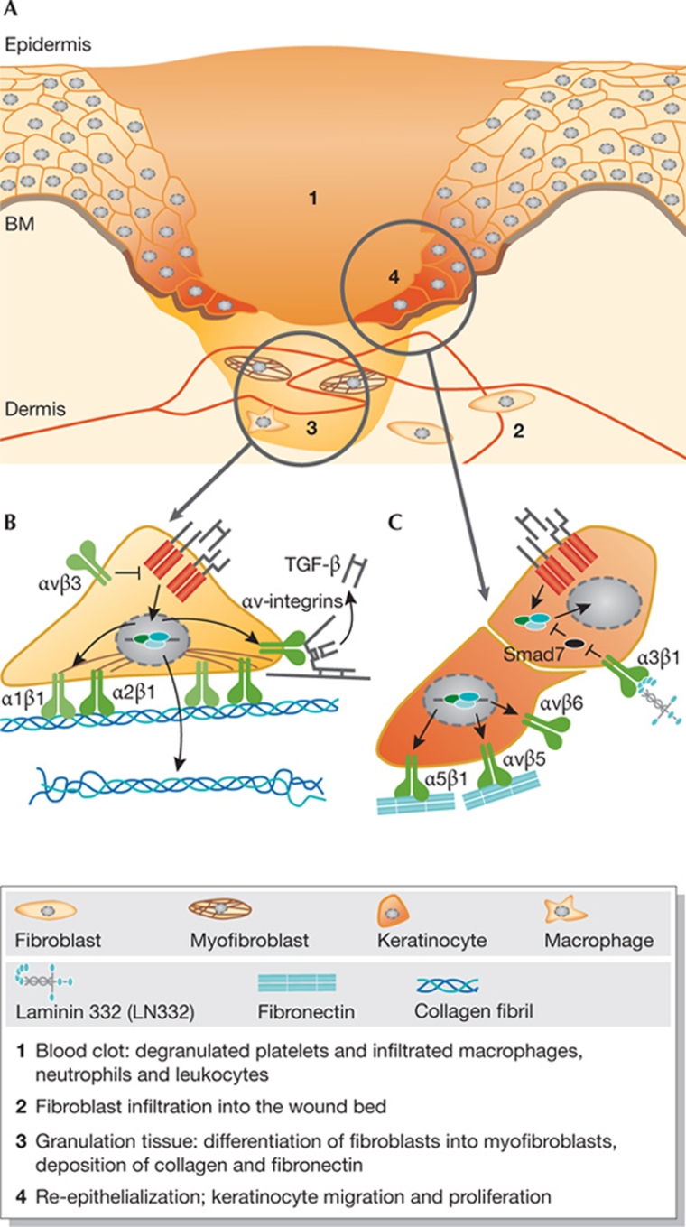
Figure 3.
Overview of proposed integrin–TGF-β interactions during wound healing. (A) Schematic representation of the main phases in wound healing, which are explained in the figure key. (B) In the granulation tissue, TGF-β induces expression of integrins α1β1 and α2β1, which mediate fibroblast contraction, and of αv-integrins, which activate latent TGF-β. Furthermore, αvβ3 might repress TGF-β signalling by inhibiting TGF-βR expression. (C) During re-epithelialization, TGF-β stimulates the expression of fibronectin and integrins, which mediate keratinocyte migration or activate latent TGF-β. Integrin α3β1 could enhance TGF-β signalling by controlling the expression of Smad7. BM, basement membrane; Col, collagen; FN, fibronectin; LN332, laminin 332.

