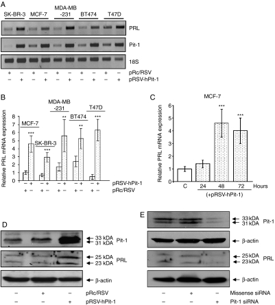
Figure 1.
Pit-1 regulates PRL expression in human mammary cell lines. (A) PRL, Pit-1, and 18S mRNA before and after 48 h of Pit-1 overexpression in the SK-BR-3, MCF-7, MDA-MB-231, BT47, and T47D cell lines. (B) Real-time PCR of PRL mRNA expression, with respect to 18S mRNA levels, evaluated in control and pRSV-hPit-1-transfected SK-BR-3, MCF-7, MDA-MB-231, BT47, and T47D cell lines. (C) MCF-7 cells were transfected with either empty pRc/RSV vector (control, C) or pRSV-hPit-1 construct at 24, 48, and 72 h respectively. Values represent means±s.d. from four independent determinations. (D) Western blots of Pit-1, PRL and β-actin in controls, and pRc/RSV (empty vector) and pRSV-hPit-1-transfected MCF-7 cells for 48 h. The major 31 and 33 kDa immunoreactive bands corresponding to Pit-1, as well as glycosylated (25 kDa) and nonglycosylated PRL (23 kDa), are indicated by arrows. (E) Western blots of Pit-1, PRL, and β-actin in control MCF-7 cells, and in cells transfected with 20 nM siRNA negative control and 20 nM Pit-1 siRNA for 48 h. (***P<0.001 and **P<0.01 versus control cells).

