
Figure 8. Role of Rac in PKA- and Epac-mediated EC barrier protection.
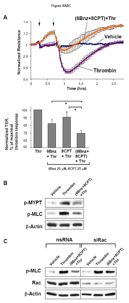
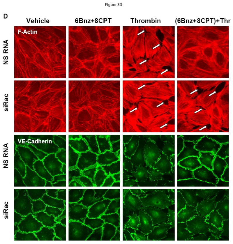
A - At the time point indicated by first arrow, cells were treated with a mixture of 6Bnz (100 μM) and 8CPT (100 μM), followed by thrombin (0.1 U/ml) challenge indicated by second arrow and measurements of TER (upper panel). Bar graphs (lower panel) represent statistical analysis of TER measurements performed in EC pretreated with either 6Bnz (25 μM), 8CPT (25 μM), or a combination of 6Bnz (25 μM) and 8CPT (25 μM) followed by thrombin challenge (0.1 U/ml, 15 min). TER decline induced by thrombin was taken as 100%; n=3-5 per condition; *p<0.05. B - Phosphorylation of MYPT1 and MLC after thrombin stimulation (0.1 U/ml, 5 min) with or without combined 6Bnz and 8CPT pretreatment was detected by western blot with specific antibodies. C - Cells were transfected with Rac-specific or non-specific siRNA for 48 hours. Effects of 6Bnz and 8CPT on thrombin-induced MLC phosphorylation were detected in control and Rac-depleted EC by immunoblotting with specific antibodies. D - Cytoskeletal remodeling was analyzed by double inmmunifluorescence staining using Texas Red phalloidin to detect F-actin and VE-cadherin antibodies to visualize adherens junctions. Paracellular gaps are marked by arrows.