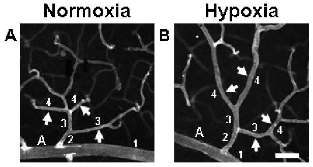
FIGURE 1.

Exposure to whole-body hypoxia resulted in an apparent increase in the diameters of arterioles and post-arteriole capillaries of murine superficial retinal microvascular networks. High magnification (20×) confocal images obtained from retinal whole-mounts labeled with lectin were used to examine hypoxia-induced changes in vessel diameter throughout the microvascular tree. Representative arterioles and capillaries (arrows) downstream of main arterioles (labeled ‘A’) exhibit increased diameters in animals exposed to systemic hypoxia (B), as compared with age-matched normoxic controls (A). To categorize vessels so that diameters could be compared for vessels of similar phenotype and relative location in the network, main arterioles were defined as vessel order 1, and sequential orders were assigned progressively downstream, incrementing at branch points at which two vessels of the same order joined (see numbers in figure) (Peirce et al., 2004). Scale bar = 50 μm.
