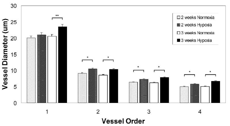
FIGURE 2.

Quantification of vessel diameters in superficial retinal microvascular networks revealed the presence of microvascular remodeling after hypoxia exposure. Vessels were categorized so that diameters could be compared for vessels of similar phenotype and relative location in the network, as demonstrated in Fig. 1. Quantification of retinal vessel diameters showed that exposure to 3 weeks of hypoxia resulted in a significant increase in the vessel diameters of main central arterioles (p = 0.003) as well as downstream arterioles (majority found to be of vessel order 2 and 3) and capillaries (majority found to be of vessel order 4) (p < 0.001). A significant increase in vessel diameters was also measured after 2 weeks of hypoxia for vessel orders 2, 3, and 4 (p < 0.001). The increase in the central arteriole diameter (vessel order 1) was not significant after 2 weeks of hypoxia exposure.
