
FIGURE 3.
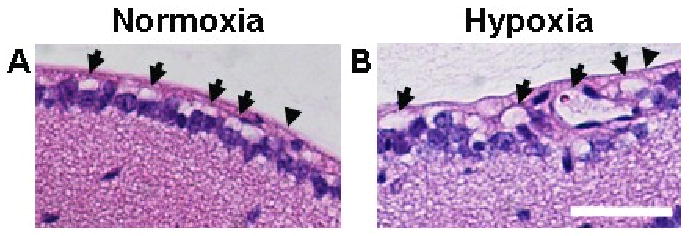
Whole-eye sections were taken from 13 wk-old mice and were stained using H&E. Whole-eye 5 μm-thick sections from mice exposed to normoxia (A) or hypoxia (B) further demonstrated diameter enlargement in vessels of the superficial retina (arrows) after 3 weeks of hypoxia exposure relative to normoxic controls. In addition, whole-eye sections indicated that the angiogenic response induced by chronic whole-body hypoxia did not involve the formation of preretinal neovascular tufts that penetrate the inner limiting membrane (arrowheads). Scale bar = 50 μm.