
FIGURE 4.
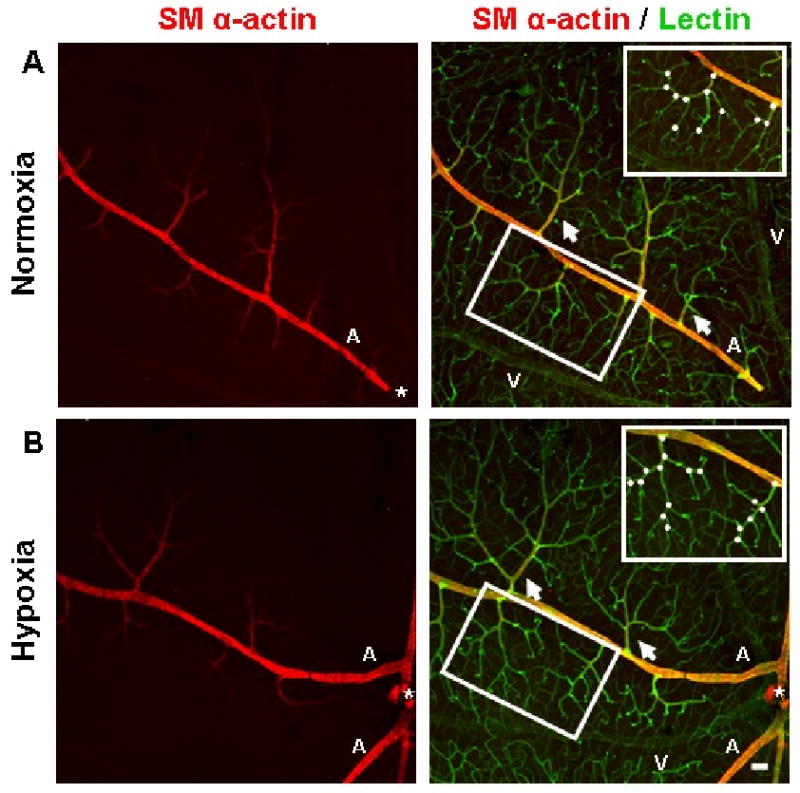
Immunohistochemistry performed on whole-mounted retinal tissue qualitatively showed that the vascular networks of the superficial retina underwent angiogenesis as a result of systemic hypoxia. Representative superficial post-arteriole vascular networks (arrows) labeled with lectin (green) from animals exposed to 3 weeks of hypoxia appear to exhibit an increased degree of vascularization (B) compared to retinal vascular networks from animals exposed to normoxia (A). Representative post-arteriole vascular networks (insets) demonstrate an increased amount of branch points (white circles) in hypoxia-exposed tissue (B) compared to networks from normoxia-exposed animals (A). Both upstream mother vessel and downstream daughter vessel were required to be located in the same confocal plane as the post-arteriole superficial network in order to be counted as a branch point. In the mouse retina, the main arterioles (labeled ‘A’) and venules (labeled ‘V’) of the superficial retina extend radially from the optic nerve (*) and are distributed alternately. Arteriole vessel identity was determined by the presence of SMA immunostaining (red). SMA investment appeared to be similar between normoxic and hypoxia-exposed retinas. Scale bar = 50 μm.