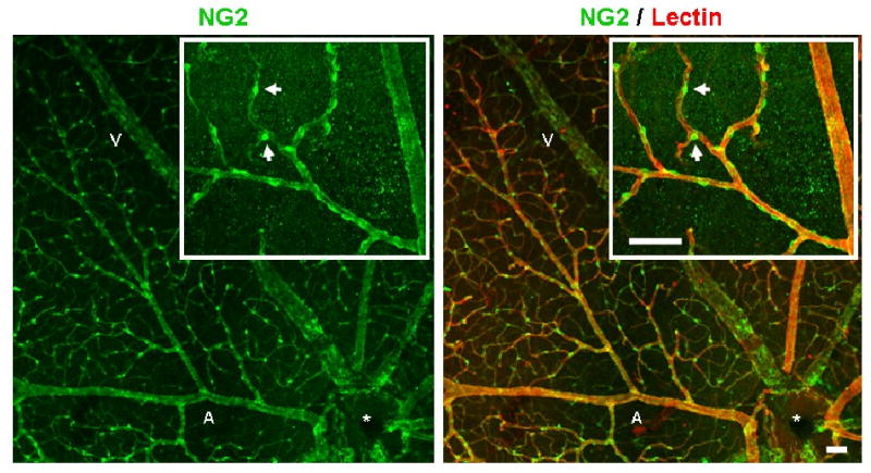
FIGURE 6.

Co-immunolabeling for pericyte marker NG2, as well as lectin, demonstrated the extensive pericyte coverage of retinal microvascular networks after 3 weeks of hypoxia exposure. All retinal microvessels labeled with lectin (red) were heavily invested by NG2-expressing pericytes (green). High magnification revealed pericyte wrapping of the main arteriole and post-arteriole capillaries (inset). Scale bar = 50 μm. Normoxia-exposed retinas displayed similar pericyte coverage density (data not shown).
