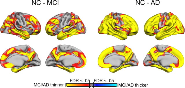
Figure 2.
Cortical thickness differences between NC and MCI/AD patients. Cortical thickness was compared point by point across the entire cortical mantle between NC and MCI patients and between NC and AD patients. Sex and age were used as covariates. The results are shown as p value maps, thresholded at FDR < 0.05. As can be seen, MCI patients have thinner cortex than NC in large cortical areas, including medial, lateral, and inferior temporal cortices, medial parietal cortex, and widespread areas in frontal cortex. The differences between NC and AD patients are even larger, covering the major part of the cortical surface, except the area around the central sulcus.

