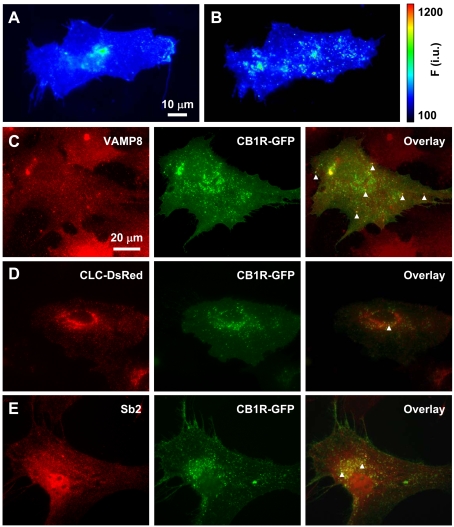
Figure 6. Intracellular CB1R localization in astrocytes.
(A and B) The presence of SEP–CB1R in low-pH intracellular structures. (A) Astrocyte expressing SEP–CB1R at rest. (B) After incubation with bafilomycin A1 to block V-ATPase, SEP–CB1R fluorescence intensity increases displaying more prominently punctate fluorescence indicating that a population of SEP–CB1R exists within acidic compartments. The pseudocolour scale is a linear representation of the fluorescence intensities ranging from 100 to 1200 intensity units (i.u.). (C–E) Intracellular CB1R–GFP localizes in endosomes. (C) CB1R–GFP (green, middle panel)-expressing astrocytes immunolabelled for endobrevin (VAMP8) (red, left-hand panel), an endosomal marker. Arrowheads mark some instances of co-localization of CB1R–GFP and endogenous VAMP8 (overlay, right-hand panel). (D) Co-expressed CLC–DsRed (red, left-hand panel), labelling early endocytic structures, and CB1R–GFP (green, middle panel) show very little co-localization (overlay, right-hand panel; arrowhead). (E) CB1R–GFP (green, middle panel)-expressing astrocytes immunolabelled for the exocytotic vesicle marker synaptobrevin 2 (Sb2) (red, left-hand panel). Subcellular localization of CB1R–GFP and Sb2 immunoreactivity show some overlap (overlay, right-hand panel; arrowheads) within the perinuclear region of the cell, whereas co-localization was not apparent at the periphery of the cell.

