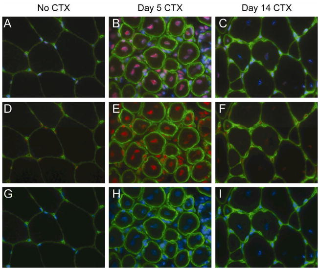
Figure 4. Mi-2 expression is up-regulated in regenerating myonuclei.
Frozen sections from uninjured and CTX-treated tibialis anterior muscles (day 5 and day 14) were mounted on a single slide and double labeled with antibodies against Mi-2 (red) and laminin (to reveal the basal lamina of individual myofibers; green). The sections were counterstained with DAPI (blue) to identify nuclei. To insure comparability, images were obtained using identical exposure settings for each section. Merged images show Mi-2, laminin, and DAPI (A–C), Mi-2 and laminin (D–F), or DAPI and laminin (G–I).

