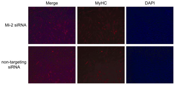
Figure 6. Knockdown of Mi-2 accelerates the formation of MyHC expressing myotubes.
48 hours after transfection with Mi-2 or non-targeting siRNA, cultures of C2C12 cells were fixed with methanol and stained with MyHC antibodies (MF20) and DAPI. The number of MHC positive tubes and DAPI staining nuclei in four random 20X fields were counted for each condition. The density of nuclei is similar in each condition. However, the cells treated with Mi-2 siRNA have a marked increase in the number of myotubes expressing MyHC (48.3 +/− 11.7 vs. 27 +/− 6.5 tubes/20X field; confidence interval 98.09%).

