Figure 10.
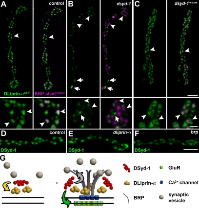
Defective DLiprin-α localization in dsyd-1 mutants. (A–C) DLiprin-αGFP/BRP-shortmStraw co-imaging in control (A), dsyd-1 (B), and dsyd-1rescue (C) are shown. The localization of DLiprin-α is changed at dsyd-1 mutant NMJs, but is rescued by reexpression of UAS–dsyd-1cDNA in motoneurons. Bars, 2 µm and 500 nm (insets). Arrowheads indicate AZs marked by BRP and arrows indicate ectopic DLiprin-α in dsyd-1 mutants. (D–F) DSyd-1 localizes to AZs in control (D), dliprin-α (E), and brp (F) animals. (G) Model of AZ assembly. Yellow arrow, DSyd-1 regulates DLiprin-α early in assembly; green arrow, DSyd-1 regulates GluR field size; gray arrow, DSyd-1 binds BRP and regulates BRP supply. Bars: (A, top): 2 µm; (A, bottom) 500 nm; (F) 2 µm.

