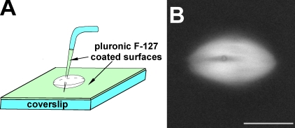
Figure 1.
Experimental approach for spindle skewering. (A) The cartoon shows the experimental setup used in all skewering experiments (see Materials and methods for more details). (B) Example of a skewered spindle visualized by the addition of X-rhodamine–labeled tubulin to the extract. The cross section of the microneedle is seen as a dark annulus. Bar, 25 µm.

