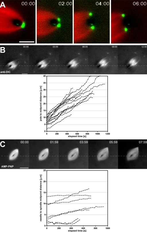
Figure 3.
Intrinsic forces push but do not pull the spindle off impaling microneedles. (A) The time-lapse series shows the dynamic morphology of a spindle pole, labeled with Alexa Fluor 488 anti-NuMA antibodies (green), as it is split by a microneedle. Spindles continued to move despite a lack of any detectable microtubules (red) on the distal side of the microneedle. (B) Time-lapse images show the behavior of skewered spindles ∼5–10 min after the addition of function-perturbing antibodies against the 70.1-kD DIC (anti-DIC). The distance between the metaphase plate and the needle were measured and plotted as a function of time for multiple spindles in the corresponding graph. (C) Assembled spindles were treated with 1.5 µM AMP-PNP and skewered within 5–10 min after treatment. AMP-PNP at this concentration inhibited flux, in agreement with nearly horizontal plots of needle to spindle midpoint distance versus time (graph). (B and C) Dashed lines indicate the position of the microneedle used to skewer the spindles. Bars: (A) 5 µm; (B and C) 25 µm.

