
Figure 1.
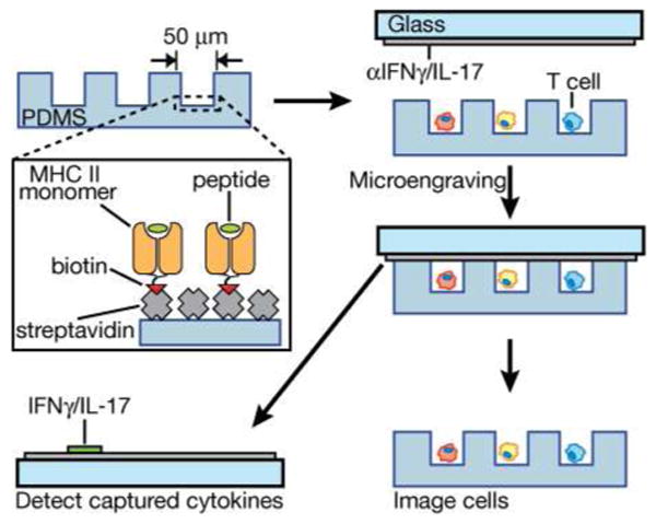
Schematic illustration of surface modification for on-chip stimulation of antigen-specific T cells and subsequent detection of released cytokines by microengraving. Streptavidin is applied to oxidized PDMS, then exposed to biotinylated monomers of recombinant, peptide-loaded MHC Class II and biotinylated anti-CD28 (not shown for clarity). The modified surface is blocked with bovine serum albumin immediately prior to loading cells. Cells are then deposited into the microwells and incubated to allow activation. After a period of time (6-18 h), the array of cells is sealed against a glass slide bearing anti-cytokine antibodies and incubated for 2 h. After this process (microengraving), the array of cells is imaged to determine the number of cells per well, and the array of captured cytokines is labeled and imaged.