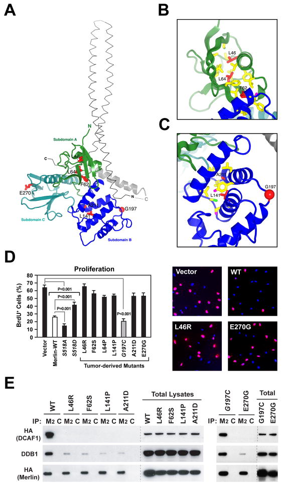
Figure 5. Several Pathogenic Missense Mutations Disrupt Binding of Merlin to CRL4DCAF1 In Vivo.
(A) Tumor derived mutants (red sticks) are shown in the context of the overall structure of the FERM domain of human Merlin (colored ribbons; PDB 1ISN). The α-helical coiled-coil domain from the crystal structure of the closed form of Spodoptera frugiperda Moesin (thin grey αcarbon trace; PDB 2I1K), which is highly homologous to human Merlin, is shown for reference. The coiled-coil domain was positioned by superimposing the FERM domains from the structures of Merlin and Moesin, which have a root mean square deviation for α-carbon atoms of 1.3 Å and share 62% sequence identity.
(B) Close-up view of tumor-derived mutations (red sticks) located in subdomain A (green ribbon) in the crystal structure of human Merlin. Residues in contact with mutated residues are drawn as sticks and colored according to atom type (carbon, yellow; sulfur, green).
(C) Subdomain B represented as in (B), with oxygens as magenta sticks. Red sphere, G197.
(D) Meso-33 cells were transiently transfected with empty vector or vectors encoding wild type Merlin or indicated mutants. Asynchronized cells growing in 10% FCS were subjected to BrdU incorporation assay. The graph indicates the percentage (± SEM) of BrdU-positive cells. Pictures show representative fields of cells transfected with indicated vectors and stained with anti-BrdU (red) and DAPI (blue).
(E) Cos7 cells were transfected with FH-tagged versions of wild type Merlin or indicated mutants. Flag- or control-immunoprecipitates (M2 and C, respectively) and total lysates were immunoblotted as indicated.
See also Supplemental Figure S5.

