Figure 3.
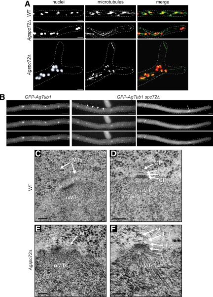
Nuclear movement, cMTs and SPB structure in Agspc72Δ. (A) Wild-type and Agspc72Δ mutants were stained with Hoechst to visualize DNA and anti-α-tubulin antibodies to detect microtubules. In the bottom image of Agspc72Δ, a cluster of nuclei can be seen at a branch site. These hyphae lack short cMTs, whereas long cMTs that extend along the growth axis are still present. A detached microtubule can be seen in the upper tip region. Bars, 5 μm. (B) Representative, deconvolved images of a Z-stack of a wild-type and two Agspc72Δ hyphae expressing GFP-AgTUB1. The complete stacks are available as Supplemental Movies S7, S8, and S9. Long and short cMTs can be seen emerging from bright foci that represent the SPBs in wild-type, but Agspc72Δ SPBs often lack associated cMTs. Arrowheads point to a long cMT in Agspc72Δ, which may facilitate bypassing. An arrow indicates an anaphase spindle. Bar, 5 μm. (C) EM image of SPB-attached microtubules in wild-type hyphae. Bar, 100 nm. The SPB is associated with nuclear microtubules (nMTs) at the IP and a tangential and perpendicular microtubule at its OP (arrows). (D) SPB structure in wild-type. Bar, 100 nm. A central plaque (CP) plus IL1 and IL2 are marked by arrows. A small amount of amorphous material could also be detected above IL1 and is part of the outer plaque (OP) (Lang et al., 2010). (E) EM image of SPB-attached microtubules in Agspc72Δ. Bar, 100 nm. The SPB is associated with nMTs at the IP and a tangential microtubule close to the central plaque (arrow). In this and other thin sections, we never observed perpendicular microtubules. (F) SPB structure in Agspc72Δ. Bars, 100 nm. A CP plus IL1 and IL2 are marked by arrows. In some cases, a small amount of amorphous material could also be detected above IL1, which could be part of the OP. However, the size of this OP remnant was substantially reduced compared with a wild-type OP (Lang et al., 2010).

