Figure 6.
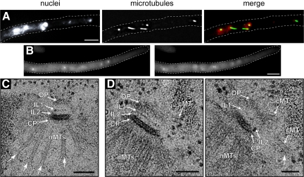
Microtubule stability and SPB structure in Agstu2Δ mutants. (A) Agstu2Δ hypha stained with Hoechst to visualize DNA and anti-α-tubulin antibodies to detect microtubules. Only short cMTs were detected. Bar, 5 μm. (B) Representative, deconvolved images of a Z-stack of an Agstu2Δ hypha expressing GFP-AgTUB1. The complete stack is available as Supplemental Movie S12. (C) EM image of a SPB from Agstu2Δ showing OP, IL2, IL1, and central plaque (CP) layers as well as nuclear microtubules (nMTs). (D) Serial sections of an Agstu2Δ SPB also show the layered SPB structure and nMTs as in A. In addition, three very short cMTs are visible, which have both perpendicular and tangential orientations with the SPB. Arrows point to “flared” microtubule ends frequently seen by EM in Agstu2Δ mutants. Bars, 100 nm.

