Figure 1.
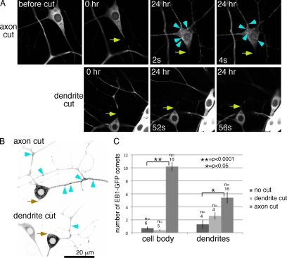
The number of growing microtubules is up-regulated by axon, but not dendrite, severing. (A) Images of EB1-GFP in the ddaE neuron were acquired before, immediately after (0 h), and 24 h after axon (top row) or dendrite (bottom row) severing. Two frames are shown from each movie from the 24-h time point. Arrows indicate the site of UV laser-mediated severing. Arrowheads point out examples of EB1-GFP comets in the cell body. In all figures dorsal is up. (B) Panels from movies acquired 24 h after axon or dendrite severing are shown. Images were inverted for ease of identifying EB1-GFP comets in dendrites; examples are marked with arrowheads. (C) The number of EB1-GFP comets in the cell body or a region of dendrite 2 was counted in single frames from movies of uninjured neurons and from neurons 24 h after axon or dendrite severing. Three frames were averaged for each animal, error bars, SD of the average from all animals. n = number of animals scored (one neuron per animal). Unpaired t tests were used to determine whether the number of comets was significantly increased after dendrite or axon cutting. No significant difference between number of dots in the cell body before and after dendrite cutting was found. Significant differences were found for cell bodies and dendrites before and after axon cutting.

