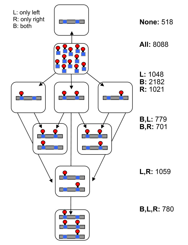
Figure 5.
Patterns in phosphorylation of adjacent phosphosites. For each pair of phosphosites (from the entire sources for phosphoproteins), the peptides that contain both of them are searched. It is then asked if from these peptides, there are peptides that contain both sites in their phosphorylated state (marked as 'both', B), only the first site is phosphorylated (marked as 'left', L) or only the second site is phosphorylated (marked as 'right', R). Each pair of sites is assigned a pattern according to the types of peptides we have seen. For example, the rightmost bar contains pairs for which we have only seen peptides in which both sites are phosphorylated (marked only with B). Note that the amount of pairs not seen in any constellation is only ~5%, indicating a high coverage of the set of experimental results that were applied for this analysis.

