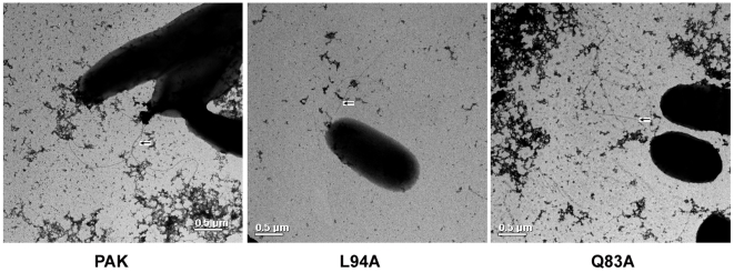
Figure 1. Electron micrographs of wild-type PAK and mutant L94A and Q83.
Bacterial were grown to OD600 = 0.5 were centrifugated at 7000 g and resuspended in PBS. A drop of bacterial suspensions was allowed to adhere to a carbon-coated grid for 10 s and drained off. Adherent cells were stained with a 0.5% uranyl acetate and examined TEM. Note the presence of flagella on the wild type and mutant bacteria. Scale bar = 500 Å.

