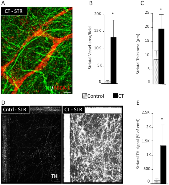
Figure 3. Increased vascular coverage and neuronal projections by angiogenic factors.
(A, B) CT treatment of organotypic slice cultures (every 4 days for 2 weeks) retains the vasculature (confocal projection for the pan-endothelial marker RECA-1 and TH), (C) increases the thickness of the striatal portion of the slice, (D,E) promotes the sprouting of TH+ fibers from the S. Nigra section to the striatal section (2-weeks after control (BSA) and CT treatment). [Size bars: 20 µm].

