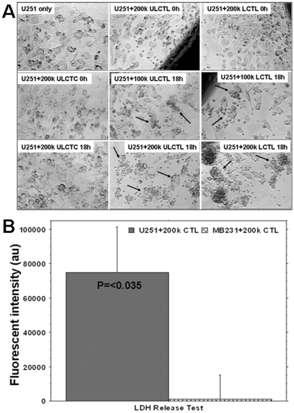
Figure 3. Specificity of labeled and unlabeled cytotoxic T-cells (LCTLs and ULCTLs, respectively) in vitro.
Figure 3A: U251 cells (100k) were plated in 24-well plates and grown until 90% confluent. Then unlabeled control T-cells (ULCTCs), LCTLs or ULCTLs were added to the cultures (100k and 200k per well). The migration and specific accumulation of T-cells around the tumor cells were microphotographed at 0h and 18h of co-culture. Left column: Morphology of U251 alone and after incubation with 200k of CTC at 0 and 18 hours. There is no change in the morphology of U251 and CTC seems to be sitting on the tumor cells even after 18 hours of incubation. Middle column: Morphology of U251 cells at 0 and 18 hours after incubation with ULCTL. The ULCTLs seem to be sitting on the tumor cells at 0 hour. The specific accumulation of ULCTLs around the tumor cells (arrows) was seen after 18 hours for both 100k and 200k ULCTLs conditions. Compared to the morphology of U251 cells at 0 hour, the dramatic changes in the morphology of U251 cells were observed. Cells become elongated and sparse. This phenomena was more pronounced at higher density (i.e. 200k) of ULCTLs. Right column: Similar to ULCTLs, LCTLs also show similar phenomena of specific accumulation around the tumor cells (arrows) and changes in the morphology of U251 cells. Figure 3B: Both U251 (100k) and human breast cancer cells (MBA-MD-231, 100k) were incubated in their respective media (1 ml in 24-well plate) in the presence or absence of 200k CTLs overnight. On the next day, supernatants from all the conditions were collected and the lactate dehydrogenase (LDH) contents were determined. Average fluorescent values detected in the supernatant of U251 and MBA-MD-231 cultures without CTLs were deducted from the fluorescent values detected in the corresponding cell lines in the presence of CTLs. The data are expressed as mean ± SEM. Significantly higher (p = <0.017) LDH activity was observed in U251 culture in the presence of CTLs indicating specificity of the sensitized T-cells.

