
Figure 1. MRI and Electroencephalogram after onset of complex partial seizures.
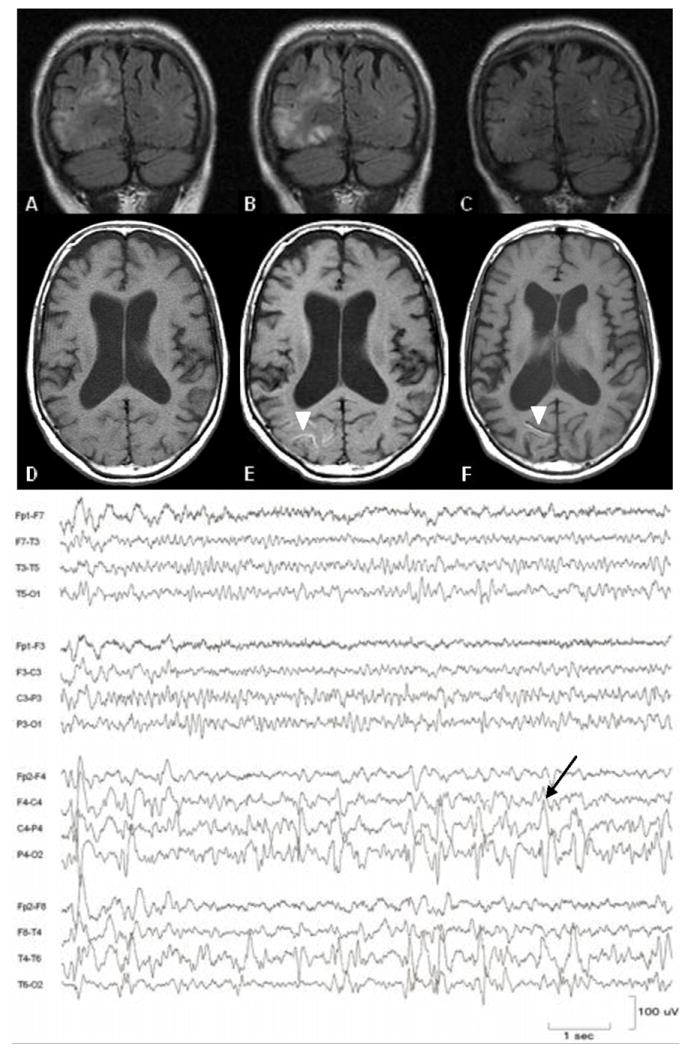
Evolution of FLAIR abnormalities (upper panel) and cortical T1 shortening (lower panel, arrowheads) one week (A, D), two weeks (B, E), and four months (C, F) after sudden onset of left hemineglect. EEG demonstrates epileptiform discharges in the right posterior quadrant, arising predominantly from the parietal region (arrow).