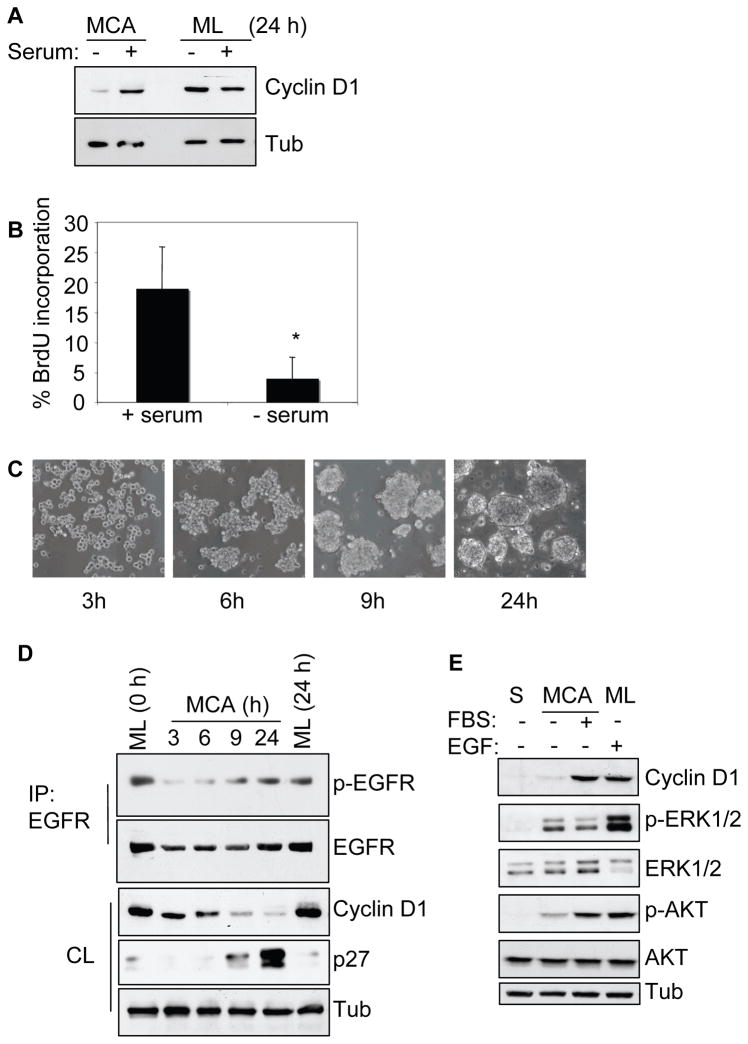
Figure 1.
Serum-deprivation suppresses cyclin D1 expression and proliferation in MCA (A) HSC-3 cells as monolayer (ML) or MCA cultured for 24 h, with or without 10% serum were lysed and analyzed by immunoblotting for cyclin D1 expression. (B) Cells subjected to MCA formation as above were assessed for BrdU uptake as described in Materials and Methods and represented as % BrdU incorporation (mean±SD, * p < 0.05). (C) Photomicrograph of HSC-3 MCAs showing the aggregate formation and compactness with time in serum-free DMEM culture. D) MCAs as in (C) were collected, lysed and analyzed for cyclin D1 and p27 expression or immunoprecipitated (IP) and analyzed for phospho-EGFR and total EGFR. Serum-starved ML cells at 0 h (time MCA started) and 24 h (time final MCA collected) were included as controls. (E) HSC-3 cells were subjected to single cell suspension (S) or as MCA in the absence or presence of serum. After 24 h, cells were collected, lysed, and processed for immunoblotting as indicated. Membranes were stripped and reprobed for total ERK1/2 and AKT. Cell lysates from serum-starved ML culture treated with EGF (10 ng/ml) for 5 min, were included as a control for ERK and AKT activation. Tubulin was used as loading control wherever indicated. The results are representatives of at least three independent experiments.

