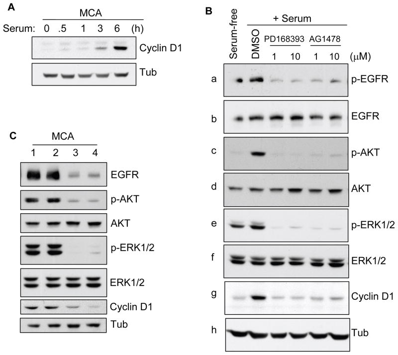
Figure 3.
Effects of EGFR signaling on serum-dependent cyclin D1 expression in MCAs. (A) HSC-3 cells were subjected to MCA formation without serum. After 20 h, aggregates (pre-formed MCA) were obtained by brief centrifugation (1000 rpm, 2 min) and incubated in 10% FBS containing DMEM in pre-coated poly-HEMA dishes. MCAs were collected at various time points as indicated, lysed, and processed for immunoblotting for cyclin D1. (B) Pre-formed MCAs as above were incubated with serum in the presence of DMSO alone, and 1 or 10 μM of PD168393 and AG1478 for 6 h. Cell lysates were prepared and immunoblotted for cyclin D1, p-EGFR, p-AKT and p-ERK1/2. Each membrane was stripped and reprobed for its corresponding total protein as indicated. Tubulin was used as an additional control protein loading. Results are representative of 2 independent experiments. (C) HSC-3 cells transfected with siRNA against EGFR were subjected to MCAs formation in presence of 10% FBS for 24 hr. Cell lysates were prepared and analyzed by Western blotting as indicated. Lane 1 is untransfected control; lane 2 is transfected with control-scrambled siRNA; lane 3 is transfected with 25 nM and lane 3 is with 100 nM EGFR-siRNA, respectively.

