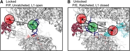
Figure 1.
Structural models of the ribosome with fluorescent components. The distances shown are in agreement with those predicted by smFRET experiments. Structural models were constructed according to published procedures (Tung et al, 2002; Munro et al, 2009a). The P-site tRNA is in blue and the L1 stalk is in purple. (A) The locked ribosome configuration with the tRNA in the classical P site, and the L1 stalk in the open position, consistent with the low-FRET (0.1) state observed in smFRET trajectories acquired from complexes with labelled L1 and P-site tRNA. (B) The unlocked ribosome configuration stabilized by EF-G (shown in cyan) in which the P/E hybrid state is formed, the L1 stalk is closed and the subunits are ratcheted—consistent with the high-FRET (0.65) state observed with labelled L1 and P-site tRNA, and the low-FRET (0.25) state observed with labelled EF-G and P-site tRNA.

