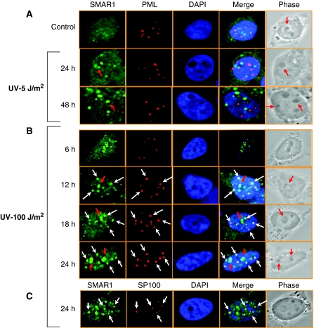
Figure 5.
Apoptotic DNA damage translocates SMAR1 into PML nuclear bodies. Immunofluorescence analysis showing SMAR1–PML co-localization in HCT116 p53+/+ cells at (A) low-dose 5 J/m2, (B) high-dose 100 J/m2 UV and (C) Co-localization of SMAR1 with Sp100 at high dose. Cells were stained with SMAR1 (green), PML (red) and Sp100 (red) antibodies and analysed by confocal microscopy. Nuclei were stained with DAPI (blue). Co-localization of SMAR1 and PML bodies are shown in white arrows. Red arrows indicate SMAR1 in nucleolus. Images shown are representative of >50 images (n>50) taken in different fields from two independent experiments.

