
Figure 1.
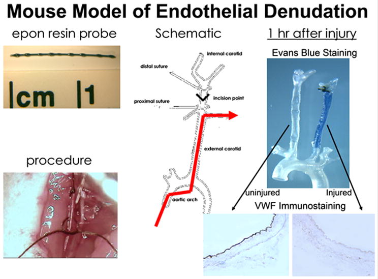
Mouse model of endothelial denudation. The top left panel shows the epon resin probe used to induce endothelial denudation of the carotid arteries in mice. The bottom left panel is a photomicrograph of the actual procedure by which the epon resin probe is inserted into the carotid artery. A schematic of the procedure is shown in the middle panel. The red line shows the restoration of blood flow after the vascular injury procedure. The right hand panel shows results of endothelial denudation 1 hr after the procedure. The top right panel is the Evans Blue staining of the left uninjured artery and the right injured artery, with the bottom right panel showing VWF immunostaining of the sections dissected from the respective carotid arteries.