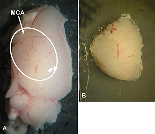
Figure 1.

Representative images of the area of multiphoton laser scanning microscopy (MPLSM) imaging (white oval) containing the cortical branches of the occluded MCA (arrow) (A) and of a 1500 µm sagittal slice glued onto a coverslip for MPLSM imaging (B).
