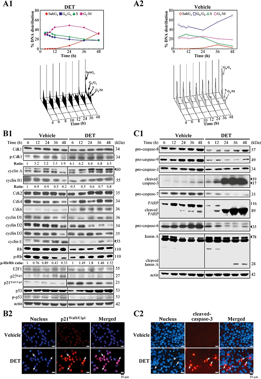
Figure 2.

DET-induced cell cycle arrest and apoptosis in TS/A cells. In (A1) and (A2), flow cytometry analysis of DNA distribution in propidium iodide-stained cells. The percentage of subG1, G0/G1, S and G2/M cells was calculated. Data are representative of three independent experiments. (B1) Immunoblotting of key biomarkers involved in G2/M phase transition in TS/A cells treated with vehicle or DET (2 µg·mL−1 at times indicated). Protein levels were normalized to the level of actin. (B2) Immunostaining of p21Waf1/Cip1 (arrows) in the nuclei of TS/A cells treated with DET or vehicle for 12 h. Cells were stained with DAPI (nuclear marker; blue) and rabbit anti-p21Waf1/Cip1 (red). (C1) Immunoblotting of apoptotic mediators in the cell lysates of TS/A cells treated with vehicle or DET. (C2) Immunostaining of cleaved form of caspase-3 (red) in TS/A cells treated with DET or vehicle for 48 h. Typical apoptotic cells (arrows) with condensed nuclei in DET-treated cells. Cdk, cell cycle-dependent kinase; DAPI, 4,6-diamidino-2-phenylindole; DET, deoxyelephantopin; PARP, poly(ADP-ribose) polymerase.
