
Figure 4.
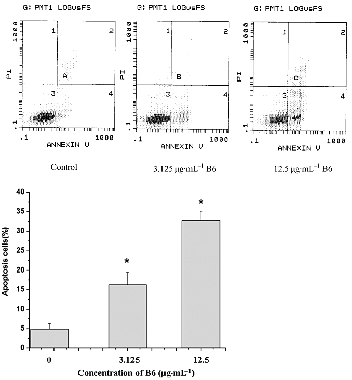
Quantification of apoptosis by AnnexinV/PI staining. HeLa cells treated with B6 for 24 h were stained with AnnexinV/PI and the % of cells undergoing apoptosis was quantified by fluorescence-activated cell sorting analysis. The values shown are the mean ± SD of data from three independent experiments. *P < 0.01 versus control group. B6, N-(4, 5-dibromo-pyrrole-2-carbonyl)-L-amino isovaleric acid methyl ester; PI, propidium iodide.