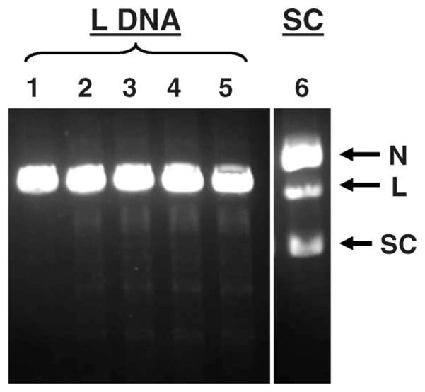
Figure 3.

Agarose gel analysis of L form of 3H-pUC19 plasmid DNA incubated with 123IEH at 4°C in PBS (pH 7.4): lane 1, control (no 123IEH); lane 2, 4.6 × 10¹² decays/ml; lane 3, 8.1 × 10¹² decays/ml; lane 4, 11.5 × 10¹² decays/ml; and lane 5, 21.9 × 10¹² decays/ml, lane 6, SC DNA exposed at the highest dose (21.9 × 10¹² decays/ml) showing DSB formation for comparison.
