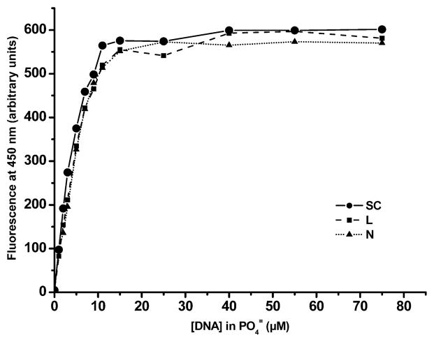
FIG. 4.
Fluorescence titration curves of supercoiled (SC), linear (L) and nicked (N) forms of pUC19 DNA with Hoechst 33342 (0.169 μM) in PBS (pH 7.4). Fluorescence maxima for all three forms of pUC19 DNA occur at approximately the same DNA concentration (20 μM), indicating saturation of ligand binding to DNA. In 125IEH–DNA incubations, the concentration of 125IEH is 284 μM, ensuring that there is little unbound 125IEH in mixtures.

