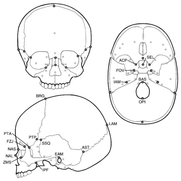
FIGURE 1.
Landmarks used in this study. All landmark locations are illustrated on each anterior, lateral, and endocranial view of the skull. Label abbreviations are from Table 2 and appear only once in this figure. Black circles represent landmarks visible in the view, and grey circles represent approximate landmark locations obscured by bone. The landmarks hormion (VSJ), hypoglossal canals (HYP), and jugular processes (JUG) are represented only by grey circles and are not labeled.

