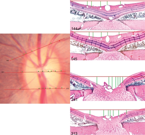
Figure 2.
Method of orienting histologic sections to the disc photograph. The topographic locations of histologic sections 145, 241, and 313 are shown. The orientation of each section (red lines in the disc photograph) is judged by assessing the relative spacing of the retinal vessels, marked by circular glyphs in the disc photograph and by vertical lines in the histologic sections. The temporal vessel outlined by a red glyph is absent in section 144, but appears in the next section (145). The superior opening in Bruch's membrane (superior disc margin) was near section 145 (described in detail in Fig. 5) and the inferior opening in Bruch's membrane was near section 335 (not shown in this figure, but described in detail in Fig. 6) Hematoxylin and eosin; magnification, ×10.

