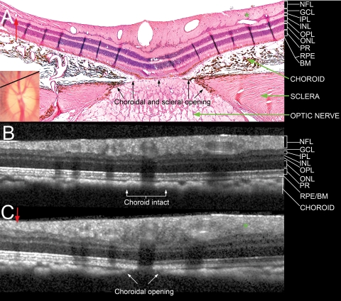
Figure 5.
Comparison of a histologic section located near the superior Bruch's membrane opening (disc margin) with matched interpolated B-scans. (A) Section 145, with its topographical location shown as a black line in the disc photograph (inset, also shown in Fig. 2). Note the presence of a small vessel in the nasal (left) part of the section (red arrow) and an oblique vessel in the temporal (right) part of the section (green asterisk). There was an opening in the choroid and sclera, but the RPE and Bruch's membrane were intact over the optic nerve. (B) An interpolated section generated from the same topographic location, at an angle of rotation of 73.5°, but with an angle of incidence of 0° (perpendicular to the surface). Neither of the vessels highlighted in (A) was detected, and the choroidal signal appeared to be continuous. (C) An interpolated B-scan generated from the same location as (B), but with an angle of incidence of −18° (angled toward the nerve). The two vessels became visible in the correct location (labeled with a red arrow and a green asterisk). An opening in the choroid was visible, the RPE/BM complex remained intact, but no signal from the deeper optic nerve and lamina cribrosa was detected. NFL, nerve fiber layer; GCL, ganglion cell layer; IPL, inner plexiform layer; INL, inner nuclear layer; OPL, outer plexiform layer; ONL, outer nuclear layer; PR, photoreceptor layer; RPE/BM, retinal pigment epithelium/Bruch's membrane complex. Hematoxylin and eosin; magnification, ×10.

