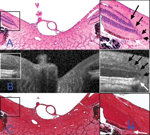
Figure 7.
Identification of the neural canal opening. Right: Magnification of highlighted regions (boxes, left). Differences in anterior surface tilt between the B-scan and the histologic sections can be explained by the presence of retinal elevation/detachment and of a rip in the retina on the left side of the sections (both features cropped out of A and C). The retinal layers in (A) and (C) are as identified in Figure 5. (A) Section 257 (hematoxylin and eosin). The termination of Bruch's membrane (the neural canal opening) is highlighted with a white arrow in the magnified view. At this location, the retinal layers taper to a point (black arrows) at the photoreceptor layer, internal to the NCO. (B) Matched interpolated B-scan. The same relationship between the retinal layers (black arrows) and the termination of the retinal pigment epithelium/Bruch's membrane complex (white arrow), as was detected in (A), are seen in the magnified view. (C) Section 260 (Ponceau S). With this stain, the distinction between the termination of the retinal pigment epithelium and an extension of unpigmented Bruch's membrane (blue arrows) was clearly visible. Magnification: (A, C) ×10.

