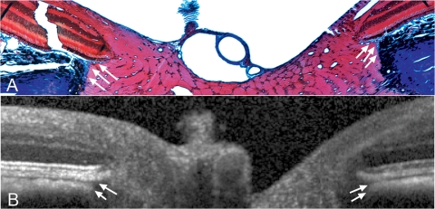
Figure 8.
Detection of border tissue of Elschnig. (A) Section 258, with border tissue highlighted with white arrows. This histologic section was located immediately adjacent to the section shown in Figure 7. The border tissue of Elschnig was seen as a connective tissue strut (stained blue) connecting the anterior sclera to Bruch's membrane and enclosing the choroid. A more complete view of this section is shown in Figure 9. Masson trichrome; magnification, ×10. (B, white arrows) The border tissue signal in the matched interpolated B-scan. SD-OCT appeared to accurately capture the orientation of the border tissue with an internally oblique configuration seen nasally (left) and an externally oblique configuration seen temporally (right).28

