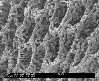
Fig. 9.

The overview of the vascular bed luminal surface of the large intestine in the scanning electron microscope. Corrosion cast. Capillaries encircling the luminal orifice of the glandulae intestinales. Bar: 100 μm. Feeding capillaries enwrap the walls and encircle the luminal gland orifice.
