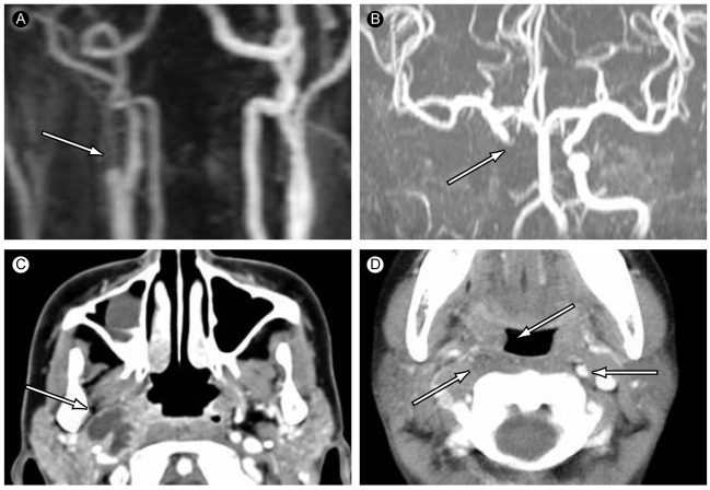
Figure 2.
Internal carotid artery flow occlusion from carotid bifurcation (A and B) due to parapharyngeal abscess propagation (C). (D) The upper white arrow shows the right cavernous sinus bulging due to inflammation, and the lower white arrow shows invisible internal carotid artery occlusion. The gray arrow shows an intact left internal carotid artery.

