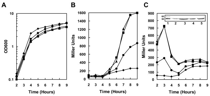
Figure 2.
Transcription analysis of pagA and atxA expression in resB, resBC and hemL mutant strains. Strains carrying a pagA-lacZ or atxA-lacZ fusion on the replicative vector pTCV-lac were grown in LB broth supplemented with kanamycin at 37°C. β-galactosidase assays were carried out on samples taken at hourly intervals as indicated. A. Cell growth of pagA-lacZ reporter strains (cell growth of atxA-lacZ reporter strains were similar) B. β-galactosidase activity of pagA-lacZ reporter strains. C. β-galactosidase activity of atxA-lacZ reporter strains. Symbols in all three panels: -◆- 34F2; -▪- 34F2△resB; -▲- 34F2△ resBC; -●- 34F2△hemL. The inset in panel C represents the Western blot analysis of AtxA on B. anthracis cell lysates collected after 3 and 8 hr of growth in LB Broth at 37°C. The amount of sample loaded on a 10% SDS-PAGE was normalized relative to cell growth. Lane 1: Magic Mark XP (Invitrogen); Lane 2: 34F2 after 3 hr of growth, Lane 3: 34F2△resB after 3 hr of growth; Lane 4: 34F2 after 8 hr of growth; Lane 5: 34F2△resB after 8 hr of growth. A full size of this Western blot is shown in Fig. S1.

