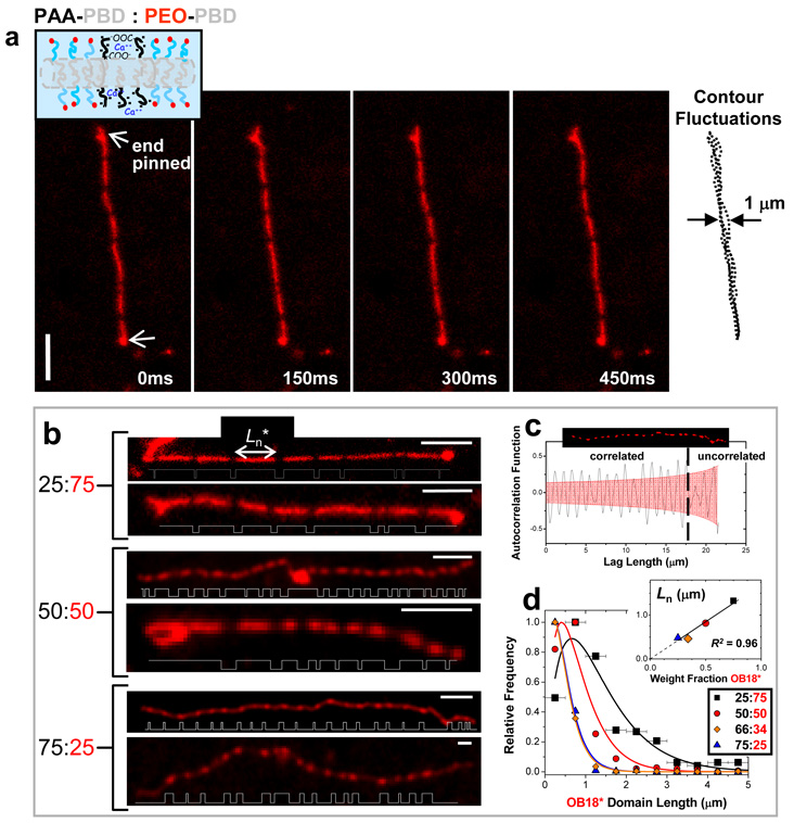
Fig. 4. Striped cylinder micelles.
(a) Time sequence of a pinned, striped worm-like cylinder micelle formed with AB1:OB18* = 25:75 at pH0 4.5 and [Ca2+] = 0.05 mM; the micelle remains flexible as evident in the contour overlay. (b) Striped cylinder micelles formed at increasing AB1 fraction, although for any sample only about 10% of cylinders appeared definitively striped with multiple micron-sized bands. Plots of thresholded fluorescence intensity along the worm contour length highlight average domain size and periodicity. (c) The long range ordering of a single 75:25 worm micelle is at least 17 µm as determined by examining the autocorrelation of the backbone fluorescence intensity. (d) Domain lengths (L) of OB18* in striped cylinders at varying AB1 fraction are fit to a Zimm-Schulz model (R2 ≥ 0.95) in which the number average length, Ln*, scales linearly (R2 = 0.96) with OB18* blend fraction (inset). Error bars represent bin sizes for L. Scale bar = 4 µm.

