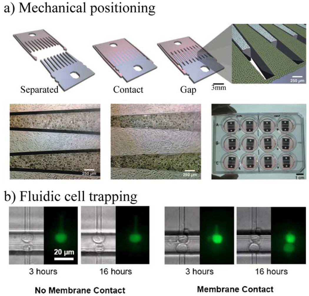
Fig. 17.
Examples of micro-scale control of cell–cell contact. (a) Micro-mechanical positioning is used to interleave cells and control cell separations with micrometer accuracy. The depicted device has three positions: fully separated, in contact, and with a small gap [91] (copyright 2007 National Academy of Sciences, USA). (b) Microchannels and fluid flow are used here to trap cells at docking sites. The docking sites can be positioned to allow cells across from one another to be in contact. The fluorescence image shows diffusion-based transfer of dye between cells only if there is membrane contact. (Reprinted with permission from [98], American Institute of Physics, 2005.)

