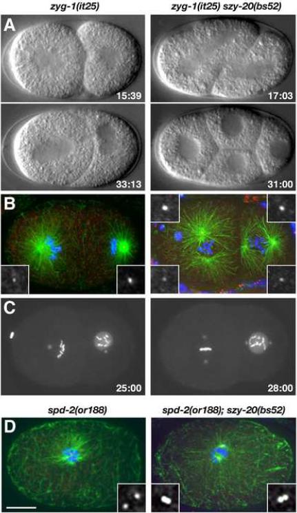
Figure 1.
Loss of szy-20 activity suppresses centrosome duplication defects in zyg-1 and spd-2 mutants. (A–C) Centrosome duplication and bipolar spindle formation are restored in zyg-1(it25) szy-20(bs52) embryos. (A) Selected images from 4D-DIC recordings. Only the double mutant embryo progresses to the four-cell stage. (B) Two-cell stage embryos stained for microtubules (green), centrioles (SAS-4, red) and DNA (blue). (C) Selected images from recordings of embryos expressing GFP-SPD-2 and GFP-histone. Time (minutes) is relative to first metaphase. (D) One-cell embryos stained for microtubules (green), SAS-4 (red) and DNA (blue). The spd-2(or188) embryo has two centrioles and the spd-2(or188) szy-20(bs52) embryo four. Each image is a Z-projection. Insets are magnified 3-fold. Bar, 10 μm.

