Figure 1.

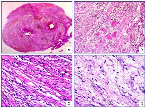
Microscopic view of IPM under low magnification (A). Cellular area formed by spindle cells and amianthoid fibers (B, C). Myxoid changes and hypocellular areas with marked edema (D) (H&E; A, ×40; B, ×200; C, D, ×400).

Microscopic view of IPM under low magnification (A). Cellular area formed by spindle cells and amianthoid fibers (B, C). Myxoid changes and hypocellular areas with marked edema (D) (H&E; A, ×40; B, ×200; C, D, ×400).