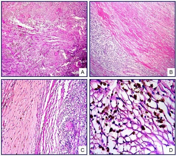
Figure 2.

IPM showing short fascicles of spindle cells and amianthoid fibers (A). The lesion contains compressed lymphoid tissue (B), intraparenchymal hemorrhage (C), and hemosiderin pigments, both free and phagocytosed by histiocytes (D) (H&E; A, ×50; B, ×50; C, ×100; D, ×400).
