
Figure 2. Mossy Fiber Sprouting.
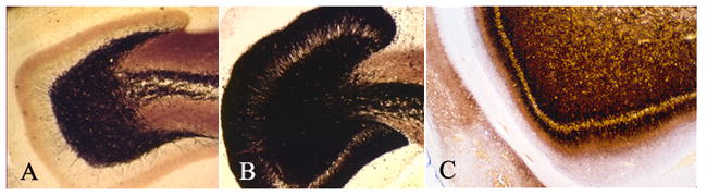
These are three photomicrographs from histological sections of the dentate gyrus (DG) stained with Timm histochemistry. Dark punctate granules depict the projection pattern of mossy fiber terminals that originate from granule cell axons. (A) The dentate gyrus from a normal rat shows dense staining in the hilus (area within the U-shape of the DG) and in the CA3 region, with an absence of dark punctate granules in the molecular layer of the DG (arrow). (B) The dentate gyrus from a rat that experienced status epilepticus induced with kainic acid demonstrates prominent staining in the molecular layer of the DG. (C) Human dentate gyrus obtained surgically during a standard anterior temporal lobectomy for the treatment of pharmacologically intractable mesial temporal lobe epilepsy. Note that the molecular layer of the DG has prominent staining demonstrating mossy fiber sprouting into that region.