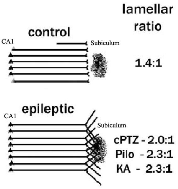
Figure 3. Synaptic reorganization in the CA1 projection to subiculum.

There is a prominent reorganization of the lamellar projection of the CA1 axonal pathway to the subiculum in several animal models of mesial temporal lobe epilepsy using retrograde tracers. In control rats, the extent of CA1 retrograde labeling from an injection site is limited to a couple of lamellas above and below of the injection site in subiculum. In contrast, in epileptic rats, the retrograde labeling extends beyond several CA1 lamellas above and below the normal projection. This is direct evidence that axonal terminals from neurons in those layers extend their axons into the area of injection (Figure modified from ref. 18).
